Extracts
Micro-Endoscope Seeks Out Early Signs Of Cancer
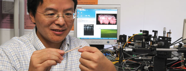
Traditional endoscopes provide a peek inside patients’ bodies. Now, a University of Florida engineering researcher is designing ones capable of a full inspection.
Physicians currently insert camera-equipped endoscopes into patients to hunt visible abnormalities, such as tumors, in the gastrointestinal tract and internal organs. Huikai Xie, an associate professor of electrical and computer engineering, is working on replacing the cameras with scanners that “see” beneath the surface of tissues — revealing abnormal groups of cells or growth patterns before cancerous growths are big enough to be visible.
“Right now, endoscopes just take pictures of the surface tissue. So, if you see some injury, or abnormality, on the surface, that’s good,” Xie said. “But most of the time, particularly with cancer, the early stages of disease are not so obvious. The technology we are developing is basically to see under the surface, under the epithelial layer.”
Experiments with Xie’s scanning “micro-endoscopes” on animal tissue have been promising, although his devices have yet to be tested in people. The pencil-sized or smaller endoscopes could one day allow physicians to detect tumors at earlier stages and remove tumors more precisely, increasing patients’ chances of survival and improving patients’ quality of life.
Xie and his graduate students have authored at least 40 papers on various aspects of the research, which is supported with more than $1 million in grants, primarily from the National Science Foundation. In September, he delivered an invited talk, “MEMS-Based 3D Optical Microendoscopy,” at the 31st Annual International Conference of the IEEE Engineering in Medicine and Biology Society. He also recently launched a small company, the Gainesville-based WiOptix Inc., to speed commercialization of his scanning technology.
With current camera-equipped endoscopes, once doctors spot abnormalities, they typically perform a biopsy, and then send the suspicious tissue to a laboratory. But biopsy is risky and may cause bleeding and even trauma. Also, it usually takes a couple of days to receive the analysis of the biopsy sample from the laboratory. If it is cancerous, surgeons may attempt to remove the abnormality and surrounding tissue, using either endoscopes equipped for surgery or traditional surgical methods.
Xie’s endoscopes replace the cameras with infrared scanners smaller than pencil erasers. The heart of his scanner is a microelectromechanical system, or MEMS, device: a tiny motorized MEMS mirror that pivots back and forth to reflect a highly focused infrared beam.
By itself, the beam strikes only a period-sized dot of tissue. But the MEMS mirror allows it to move meth-odically back and forth, scanning a fingernail-sized piece of tissue row by row, like a lawnmower moving across a yard. The resulting image is high resolution: Xie said his scanners have achieved resolution of 10 microns, or 10 millionths of a meter, in laboratory tests. That’s more than 10 times higher resolution than the only other non-camera-based endoscopes on the market, which use ultrasound technology, he said. The high-resolution image also includes depth information, so the risky biopsy can be more specific to avoid randomness, or even completely avoided.
Computers process the return signal from the endoscopes, transforming it into a three-dimensional image of the surface tissue and the tissue beneath. One scanner even produces a 360-degree image of all the tissue surrounding the endoscope. Doctors or other trained observers can then search the image for abnormalities or suspicious growth patterns.
Xie said doctors could use the endoscopes not only for diagnosis but also for treatment and surgery. Currently, he said, doctors must rely during operations on static MRI or CT images of tissue obtained before the operation begins. But his scanners make images available in real time. That would be particularly useful for regions of the body where removing as little tissue as possible is paramount, for example in brain surgery, he said.
“We are trying to couple this imaging probe with cutting tools, so that when surgeons begin cutting, they know exactly what’s in front of them,” he said.
David Dickensheets, a professor of electrical and computer engineering at Montana State University in Bozeman, said Xie’s research shows great potential.
“The impact on quality of care could be huge, allowing more comprehensive screening than is possible with point biopsies, and making it possible to achieve both diagnosis and treatment in a single patient visit,” he said in an e-mail. “Professor Xie’s research is at the leading edge of this emerging technology area, and he has worked hard to move the technology out of the laboratory and into demonstration instruments that show its potential.”