Extracts
Imaging Solves Brazilian Horse's Sinus Headache
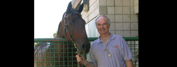
After surviving an odyssey of complicated medical problems and difficult surgeries, a Brazilian Olympic dressage horse named Livello is recuperating back in his home country, thanks to University of Florida veterinarians.
This is a horse that came all the way from Brazil because we had the technology to treat him,” said UF equine surgeon David Freeman.
Freeman said Livello’s case illustrated the importance of powerful imaging equipment such as UF’s MRI unit in guiding effective medical treatment.
“Livello actually came here because the owners were aware we had CT and thought that could be used to help him, but it turned out that the MRI was a better imaging tool for his problem,” Freeman said.
Livello’s story began in Brazil last October with a bad tooth. A tooth extraction procedure damaged the horse’s tear duct and intraorbital nerve, veterinarians said.
“Tears were coming down his face, and he had nerve damage that was causing him to rub his face and sneeze,” Freeman said, adding that a subsequent procedure did not resolve the problem. “The surgeries went well, but never cleared up the infection Livello had developed in his sinuses.”
Because of the infection, Livello subsequently developed facial swelling and a malodorous nasal discharge.
Livello’s owners and their veterinarians had heard of Freeman and UF’s imaging capability and decided to bring the horse to Gainesville. In February, owner Jorge de la Rocha, who also has ridden Livello as part of the Brazilian Olympic dressage team, flew the horse and veterinarian Patricia Brossi to UF’s Alec P. and Louise H. Courtelis Equine Hospital.
“We had some idea based on Livello’s history and clinical signs that there was probably some necrotic bone that needed to be removed,” Freeman said. “But we didn’t know the exact location or extent of it, and that is where both the CT and our new MRI unit came in.”
An initial surgery resulted in the removal of a lot of dead bone and tissue, but Livello’s sinus drainage continued, as did the nasal discharge.
“So we did another MRI on him about three weeks later and then another surgery after that,” Freeman said. “The MRI images helped us find the sites where we needed to go, and the site was not an easy area to gain access to.”
Within weeks of the third surgery, however, Livello’s nasal discharge had vanished.
“Every now and then we get cases that test us and test our general ability to handle very serious veterinary challenges, and this was one of them,” Freeman said.